All published articles of this journal are available on ScienceDirect.

Chronic Recurrent Multifocal Osteomyelitis (CRMO) in Disguise: A Case Report
 iD
Girish Chandra Bhatt1
iD
Shikha Malik1
iD
Authors Info & Affiliations
Abstract
Background
Chronic Recurrent Multifocal Osteomyelitis (CRMO) is an uncommon inflammatory bone disorder in children, characterised by sterile lesions and a relapsing course. Because its presentation overlaps with infection and malignancy, diagnosis is often delayed, especially in regions where tuberculosis is prevalent.
Case Presentation
A 12-year-old boy presented with a 3-month history of intermittent fever, progressive lower-limb pain, and difficulty in walking. The illness was marked by migratory joint involvement and weight loss. Examination showed localised tenderness and restricted joint movement. Laboratory evaluation revealed anaemia and elevated inflammatory markers, while microbiological and autoimmune investigations were negative. Imaging demonstrated erosive and mild osteosclerotic changes on radiographs and multifocal marrow oedema on whole-body MRI involving the femora, pelvis, and sacroiliac joints without abscess or bone destruction. Bone marrow examination excluded infection and malignancy. Based on clinical and radiological findings, this study made a diagnosis of CRMO. Initial treatment with non-steroidal anti-inflammatory drugs provided partial relief. Because the disease persisted, corticosteroids and methotrexate were introduced, resulting in marked clinical improvement and recovery of mobility. At 6 months of follow-up, the child remained asymptomatic, with improvement of inflammatory markers and no further relapse.
Conclusion
CRMO should be considered in children with chronic bone pain and sterile inflammatory findings. Early recognition and appropriate imaging are essential to avoid misdiagnosis and prevent long-term complications. Whole-body MRI is valuable for demonstrating multifocal disease and supporting early diagnosis, thereby avoiding unnecessary antimicrobial therapy and invasive investigations.
1. INTRODUCTION
Chronic Recurrent Multifocal Osteomyelitis (CRMO) is a non-infectious, immune-mediated bone disorder that affects children and adolescents, with females more commonly affected. Although first described more than five decades ago, it remains underrecognized because of its protean manifestations and the lack of definitive diagnostic tests [1]. CRMO typically presents with insidious bone pain, often without fever or overt systemic symptoms, and may mimic a wide range of conditions, including infectious osteomyelitis, malignancies, such as Ewing sarcoma, or Langerhans cell histiocytosis (LCH) [2].
The diagnostic dilemma is particularly heightened in TB-endemic regions, where lytic bone lesions are frequently attributed to skeletal TB. In this context, reliance on clinical criteria and advanced imaging, particularly whole-body MRI (WB-MRI), has become paramount. WB-MRI offers a sensitive, non-invasive method for detecting characteristic bone marrow oedema and multifocal involvement, thereby facilitating diagnosis and monitoring without biopsy in typical cases [3].
Herein, this study presents a diagnostically challenging case of CRMO in a young male child from a TB-endemic region whose multifocal bone involvement and systemic features necessitated a multidisciplinary, exclusion-based approach.
2. CASE DESCRIPTION
A 12-year-old male presented with a 2-3-month history of intermittent low-grade fever and progressively worsening lower-limb pain. The symptoms began insidiously following a trivial fall from stairs, after which he developed dull, non-radiating pain over the left hip and thigh. The pain worsened with movement and was partially relieved with rest and analgesics. Over subsequent weeks, joint movement progressively restricted, with swelling initially involving both hips and the left thigh, later extending to the left knee and ankle, with associated warmth and tenderness. Weight-bearing became increasingly difficult, first affecting the left lower limb and later involving the right hip, knee, and ankle joints. Interestingly, as the right-sided symptoms progressed, the left-sided complaints partially resolved.
The fever remained intermittent and low-grade, without chills, rigours, or any identifiable infectious focus. There were no associated respiratory, cardiac, neurological, or gastrointestinal symptoms. The child had significant weight loss and reduced appetite. There was no history of similar illness, chronic medication use, or prior hospitalisations.
On examination, the child was alert and haemodynamically stable, with pallor noted. There was no clubbing, lymphadenopathy, or peripheral oedema. He preferred a supine posture with the right lower limb mildly flexed and externally rotated. Local examination revealed tenderness and mild swelling over the right thigh with painful restriction of movements at the hip, knee, and ankle joints; hip flexion was limited to approximately 110°. Mild tenderness and restricted range of motion were also noted over the lower back and bilateral hips. Systemic examination was otherwise unremarkable.
Laboratory evaluation demonstrated microcytic hypochromic anaemia, markedly elevated ESR and CRP, and thrombocytosis. Iron studies showed low serum iron with reduced transferrin saturation, suggesting iron-restricted erythropoiesis, while vitamin B12 and folate levels were normal. The raised acute-phase reactants and thrombocytosis supported the presence of an active systemic inflammatory process but were nonspecific and therefore could not distinguish CRMO from chronic infection or malignancy. Liver and renal function tests were within reference ranges. Autoimmune screening was negative, blood cultures were sterile, and subsequent microbiological investigations for tuberculosis were negative (Table 1).
| Investigation | Result | Reference Range |
|---|---|---|
| Hemoglobin | 8.9 | 12-16 g/dl |
| WBC Count | 9400 | 4000 - 12000/cu.mm |
| Platelet Count | 611000 | 150000-450000/cu.mm |
| ESR | 96 | <10 mm 1st hour |
| CRP | 42 | <5.00 mg/L |
| Total Calcium | 10.60 | 8.8 - 11 mg/dl |
| Phosphate | 5.59 | 2.5 - 4.5 mg/dl |
| Vitamin D | 42.6 | 30 - 80 ng/ml |
| Urea | 15.12 | 20-40 mg/dl |
| Creatinine | 0.52 | 0.6 - 1.2 mg/dl |
| ANA | Negative | |
| RA Factor | Less than 9.62 | < 10 IU/ml |
| C3 | 1.9 | 0.9-1.8 g/L |
| ASO | 55.6 | <200IU/ml |
| Bone marrow Study | Reactive Marrow No Blasts noted. | |
| Bone Marrow culture | Sterile | |
| Gastric Aspirate & Bone marrow CBNAAT | Negative | |
Plain radiographs of the hip joints revealed erosive and mild osteosclerotic changes (Fig. 1). Whole-body MRI demonstrated multifocal marrow signal abnormalities involving the bilateral lower limbs and pelvis. A well-defined metaphyseal lesion in the left distal femur extended across the physis into the epiphysis, appearing T1 hypointense and T2 fat-suppressed hyperintense, consistent with marrow oedema. Similar but less extensive lesions were noted in the right distal femur. Additional periphyseal marrow oedema involved the greater trochanters, left ischiopubic ramus, acetabular roof near the triradiate cartilage, and right sacroiliac region, with adjacent soft-tissue oedema but no abscess (Fig. 2).
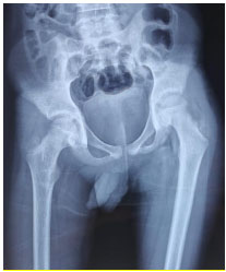
Anteroposterior radiograph of the pelvis showing erosive and mild osteosclerotic changes around bilateral hip joints.
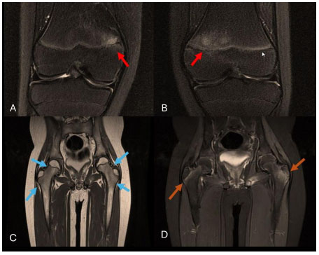
A. MRI (T2 sequence with fat suppression) Right knee showing T2 FS Hyperintense, T1 Hypointense altered marrow signal (red arrow) in the medial aspect of the distal metaphysis of the right femur. B. T2 FS Hyperintense altered marrow signal (red arrow) seen in the medial aspect of the distal metaphysis of the left femur. C&D. showing T2 FS Hyperintense altered marrow signal (blue & brown arrows) seen adjacent to the growth plate of the bilateral greater trochanter and femoral neck-shaft junction (left>right).
Bone marrow examination and microbiological studies were negative for malignancy and TB. According to the Jansson classification framework, the patient fulfilled two major criteria: radiologically demonstrated osteosclerotic bone lesions and multifocal skeletal involvement. The elevated ESR and CRP further supported a minor inflammatory criterion. The duration-of-observation criterion was not considered fulfilled because symptoms had been present for only 2-3 months at presentation, and a sterile inflammatory bone biopsy was not counted because a targeted bone-lesion biopsy was not performed.
Initial treatment with NSAID resulted in partial symptomatic improvement. However, because of persistent/recurrent disease activity, Prednisolone was initiated at 2 mg/kg/day orally for 4 weeks, followed by a 4-week tapering regimen. Subcutaneous methotrexate 15 mg/m2 once weekly was subsequently initiated, with folic acid supplementation. Clinical improvement was evident within 24 weeks, with progressive reduction in bone pain and joint swelling and restoration of independent ambulation. At the latest follow-up of 6 months, the child remained asymptomatic, with no further relapses. Haemoglobin improved from 8.9 to 13 g/dL, ESR from 96 to 10mm/h, and CRP from 42 to 8 mg/L. A follow-up WB-MRI/radiography is planned at 12 months. Consent was obtained from the patient and parents for publication.
3. DISCUSSION
CRMO is a rare, non-infectious, autoinflammatory skeletal disorder. It affects children and adolescents. First described in 1972 by Giedion et al., CRMO has since evolved from an obscure clinical entity into a recognised prototype of autoinflammatory bone disease. Despite this, its diagnosis remains elusive and is often delayed—particularly in low-resource or TB-endemic settings, where infectious and neoplastic mimickers are more routinely considered [4].
CRMO is an autoinflammatory bone disorder driven predominantly by innate immune dysregulation rather than infection or autoantibody-mediated mechanisms. Although its precise pathogenesis remains incompletely understood, an imbalance between pro- and anti-inflammatory cytokines appears central. Dysregulated monocyte signaling and inflammasome activation promote increased production of IL-1β, IL-6 and TNF-α, with relative impairment of anti-inflammatory cytokines such as IL-10. These changes enhance RANK-RANKL-mediated osteoclast differentiation and activation, contributing to sterile bone inflammation, osteolysis and subsequent remodelling. Genetic susceptibility is also recognised [1, 2]. Rare monogenic CRMO-like autoinflammatory disorders include LPIN2-related Majeed syndrome and IL1RN-related deficiency of the IL-1 receptor antagonist, while variants in FBLIM1 and FGR have been implicated as susceptibility factors in some patients with sporadic CNO [1, 2]. Nevertheless, most childhood CRMO remains genetically complex, and no single causative genetic abnormality has been identified in most patients.
The rarity of CRMO (incidence ~1 in 1,000,000) hinders early recognition. Its nonspecific presentation includes intermittent bone pain, joint swelling, and low-grade fever. These symptoms closely mimic infectious and malignant conditions. This often results in misdiagnosis, unnecessary antibiotic therapy, and avoidable invasive diagnostic procedures [5]. The patient, a 12-year-old male with progressive pain and swelling of the lower limb joints, initially raised concern for skeletal TB.
In this context, ruling out mimickers becomes critical. Infectious osteomyelitis typically presents with an acute onset, elevated white cell counts, local erythema, and purulence. It often has a single bone focus. In contrast, CRMO normally evolves subacutely. It usually presents with multifocal, symmetric lesions in the metaphyses of long bones. TB, particularly skeletal TB, is characterised by systemic signs, including weight loss, night sweats, and caseating granulomas. Radiologically, it may present with vertebral body collapse, paraspinal abscesses, or sinus formation. These features were absent in the patient. All microbiologic investigations, including blood cultures, bone marrow aspirate cultures, CBNAAT (GeneXpert-MTB/RIF), and AFB staining, were sterile. This lack of microbial evidence, combined with chronicity, favoured the diagnosis of CRMO [3, 5].
Differentiation from malignancies, such as Ewing sarcoma and LCH, is crucial. Ewing sarcoma often presents with systemic symptoms like fever and weight loss. It also shows a permeative pattern of bone destruction and periosteal reaction on radiographs. LCH may present with punched-out lytic lesions, soft-tissue masses, and diabetes insipidus in its multifocal form. In contrast, the absence of periosteal reaction, lack of systemic toxicity, and multifocal marrow oedema without cortical destruction on MRI supported a non-malignant inflammatory pathology [4, 6].
Advanced imaging, particularly WB-MRI, has revolutionised the diagnosis of CRMO. WB-MRI is non-invasive, radiation-free, and enables early detection of both symptomatic and asymptomatic lesions. It remains the gold standard in suspected CRMO cases [3]. In this patient, WB-MRI revealed multiple bilateral metaphyseal and periphyseal lesions involving the distal femora, pelvis, and sacroiliac joints. There was no abscess, periosteal reaction, or bone destruction. These findings closely mirrored the classic topography described in CRMO cohorts [7].
Previous imaging studies have shown that commonly affected sites include the metaphyses of the long bones, the pelvis, the clavicle, and the vertebral bodies, with bilateral and periphyseal involvement representing characteristic imaging patterns [3,7]. In the current case, the symmetric involvement of the weight-bearing bones adjacent to the growth plates further supported the diagnosis. Notably, growth-plate involvement posed a dilemma regarding biopsy. In keeping with best practices and recent literature, biopsy was deferred to avoid disrupting skeletal growth, particularly since malignancy and infection had been excluded [4, 6].
Several diagnostic frameworks have been proposed for CNO/CRMO, although no universally accepted gold-standard diagnostic test exists. The Jansson criteria combine major features—including radiologically demonstrated osteolytic or sclerotic lesions, multifocality, palmoplantar pustulosis/psoriasis and sterile inflammatory bone biopsy—with minor clinical and laboratory features. In contrast, the Bristol criteria place greater emphasis on typical clinical and radiological findings and use the extent of skeletal involvement and CRP concentration to determine the need for biopsy. Multifocal disease (or isolated clavicular involvement) with typical imaging and CRP <30 mg/L can support diagnosis without biopsy, whereas unifocal non-clavicular disease or CRP ≥30 mg/L generally warrants biopsy to exclude infection and malignancy. Thus, both approaches emphasise characteristic imaging and exclusion of alternative diagnoses, while the Bristol criteria provide a more pragmatic framework for determining when histopathological confirmation is required. Two major Jansson criteria were fulfilled in this patient: radiologically demonstrated osteosclerotic bone lesions and multifocal skeletal involvement. The characteristic distribution of lesions on whole-body MRI, together with negative microbiological and malignancy evaluations, further supported the diagnosis of CRMO [8].
Management of CRMO is stepwise and depends on disease severity and response to first-line agents. Non-steroidal anti-inflammatory drugs (NSAIDs) are the mainstay of initial treatment and are effective in up to 60% of cases [6]. In our case, NSAIDs were promptly started and produced early clinical improvement. However, the presence of multifocal pelvic involvement placed the child at risk for recurrence and progression. Because of persistent/recurrent disease activity despite NSAID therapy and multifocal pelvic involvement, treatment was escalated to corticosteroids followed by methotrexate. DMARDs are used in patients with poor response to NSAIDs or high-risk lesions (e.g., pelvic or spinal). Refractory or axial disease may require TNF-α inhibitors such as etanercept or adalimumab. Studies have demonstrated their efficacy in inducing remission and preventing vertebral collapse or deformities [9]. For vertebral or severe osteolytic lesions, bisphosphonates may be considered for their anti-resorptive and anti-inflammatory effects [9, 10].
Long-term monitoring is essential, given the chronic relapsing-remitting nature of CRMO. Even in clinically asymptomatic children, imaging may reveal ongoing or new lesions. Menashe et al. emphasised that subclinical activity may persist and contribute to long-term structural compromise if not adequately monitored [7]. WB-MRI enables baseline assessment and facilitates serial follow-up and objective evaluation of treatment response. Follow-up WB-MRI or radiography was not available at the 6-month assessment; therefore, radiological resolution or persistence of subclinical lesions could not be evaluated despite the marked clinical and biochemical improvement. Repeat imaging is planned at 12 months.
This report has certain limitations. As a single-case observation, its findings may not represent the full clinical spectrum of CRMO. Histopathological confirmation of the bone lesions was not obtained; however, the characteristic multifocal imaging pattern and exclusion of infection and malignancy supported the diagnosis. In addition, the relatively limited follow-up restricts assessment of long-term disease activity, relapse, and treatment response.
However, multidisciplinary care is essential, with early involvement of paediatric rheumatology, radiology, orthopaedics, and physiotherapy to optimise outcomes. Family counselling helps set expectations for disease course and adherence. Early physiotherapy and structured follow-up in our patient aided mobility and prevented joint contractures.
CONCLUSION
This case emphasises the importance of early recognition of CRMO in avoiding inappropriate antimicrobial use, invasive procedures, and delayed therapy. In high TB-burden settings, clinicians must remain vigilant and integrate clinical, radiologic, and laboratory parameters to differentiate CRMO from its mimickers. With increased awareness and systematic evaluation, early diagnosis and appropriate treatment can prevent permanent damage and significantly improve the long-term quality of life in affected children.
AUTHORS’ CONTRIBUTIONS
The authors confirm their contributions to the paper as follows: MD contributed to the conception and design of the study, the acquisition and analysis of data, the interpretation of data, and the drafting of the manuscript. SR contributed to data acquisition and manuscript drafting. JS contributed to the analysis and interpretation of data and the critical revision of the manuscript for important intellectual content. AK contributed to data acquisition and manuscript revision. GCB contributed to the study design and critical revision of the manuscript. SM supervised the study, contributed to the interpretation of data, and critically revised the manuscript for important intellectual content. All authors approved the final manuscript.
LIST OF ABBREVIATIONS
| AFB | = Acid-Fast Bacilli |
| ANA | = Antinuclear Antibody |
| ANCA | = Antineutrophil Cytoplasmic Antibody |
| CBNAAT | = Cartridge-Based Nucleic Acid Amplification Test |
| CNO | = Chronic Nonbacterial Osteomyelitis |
| CRMO | = Chronic Recurrent Multifocal Osteomyelitis |
| CRP | = C-Reactive Protein |
| DMARDs | = Disease-Modifying Antirheumatic Drugs |
| ESR | = Erythrocyte Sedimentation Rate |
| LCH | = Langerhans Cell Histiocytosis |
| MRI | = Magnetic Resonance Imaging |
| MTB/RIF | = Mycobacterium tuberculosis/Rifampicin |
| NSAIDs | = Non-Steroidal Anti-Inflammatory Drugs |
| TB | = Tuberculosis |
| WB-MRI | = Whole-Body Magnetic Resonance Imaging |
ETHICS APPROVAL AND CONSENT TO PARTICIPATE
Ethical approval was waived by the Institutional Ethics Committee as this is a single case report. Written informed consent was obtained from the patient’s parents/legal guardians for participation.
HUMAN AND ANIMAL GUIDELINES
This study was conducted in accordance with the ethical principles outlined in the Declaration of Helsinki. No animal subjects were involved in this study.
CONSENT FOR PUBLICATION
Written informed consent was obtained from the patient’s parents/legal guardians for publication of this case report and any accompanying images. All efforts have been made to maintain patient confidentiality and anonymity.
ACKNOWLEDGEMENTS
Declared none.