All published articles of this journal are available on ScienceDirect.

Wiskott-Aldrich Syndrome Mimicking Relapsing Polychondritis in a Middle-aged Woman: A Case Report
 iD
Authors Info & Affiliations
Abstract
Background
Inborn errors of immunity pose a diagnostic challenge for rheumatologists, as they can masquerade as various autoimmune diseases. Maintaining a low threshold for investigation is crucial to prevent delays in diagnosis. Additionally, the atypical presentation of an X-linked disorder in a female patient is unlikely and leads to a late diagnosis and suboptimal treatment; thus, our case report aimed to raise awareness of these complex and often overlooked conditions.
Case Presentation
Herein, we have described the rare case of Wiskott-Aldrich syndrome, an X-linked disease, presenting in a middle-aged woman who was initially diagnosed with relapsing polychondritis. By reviewing the patient’s medical and family history, we identified key clues of immunodeficiency, a low platelet count, and a son with features consistent with Wiskott-Aldrich syndrome, which could have prompted earlier testing and recognition of her condition.
Conclusion
While autoimmune diseases may resemble inborn errors of immunity, a cautious examination of the patient’s familial history and atypical diagnostic odyssey may help discriminate between the diagnoses.
1. INTRODUCTION
Wiskott-Aldrich syndrome (WAS) is a rare X-linked disease due to the WAS mutation. It mainly affects male patients and encompasses different clinical phenotypes. It is usually characterized by eczema, microthrombocytopenia, immunodeficiency, and autoimmune manifestations [1], among which inflammatory arthritis occurs in 29% of the cases [2]. As an X-linked syndrome, it rarely presents in female patients, and if so, it harbors incomplete or unusual characteristics.
2. CASE PRESENTATION
Herein, we have described the case of a 51-year-old woman who was first evaluated in her late twenties for the development of ankles’ arthritis, purpura, and bilateral alternating anterior uveitis. Skin punch biopsy revealed non-specific leucocytoclastic vasculitis. Over the next years, she gradually developed right ear and nose chondritis, right neurosensorial deafness, relapsing anterior scleritis, and peripheral neuropathy (Fig. 1). This combination of manifestations led to the provisional diagnosis of relapsing polychondritis. Her past medical history was noticeable for immune thrombocytopenia (ITP) and the early death of her male child at the age of 2 months with fever, thrombocytopenia, and hepatosplenomegaly, while she was in her early twenties. No genetic testing was available, and this information was initially deemed irrelevant to her clinical presentation.
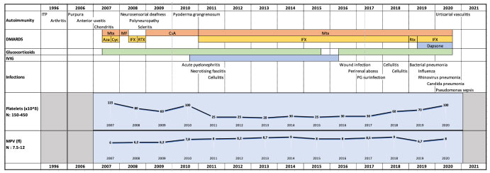
Visual representation of the evolution of the patient’s condition and treatments.
Four years later, her autoimmune profile got more complicated by several infections that were attributed to her immunosuppressive regimen. She was admitted for large-scale and refractory pyoderma gangrenosum (PG) lesions on her lower legs and buttocks (Fig. 2). This prolonged hospitalization was complicated by acute pyelonephritis and extensive necrotizing fasciitis of the abdomen, perineum, and right thigh. Considering multiple treatment failures for her PG lesions and the infectious episodes, intravenous immunoglobulin (IVIG) treatment was empirically added (2g/kg) every 4 weeks. This treatment, in addition to infliximab, methotrexate, and low-dose prednisone, improved her PG lesions and controlled her infectious propensity.
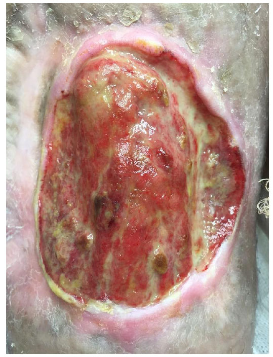
Extensive pyoderma gangrenosum lesion of the lower leg.
Five years later, shortly after the IVIG treatment was weaned, the patient’s bacterial, fungal, and viral infections relapsed (from 2016 to 2020), which included multiple wound infections (including infection of her PG lesions); perirenal abscess; multiple instances of cellulitis; bacterial pneumonia; viral pneumonia secondary to rhinovirus, enterovirus, and influenza; infectious pulmonary nodules secondary to Candida and bacteremia due to Pseudomonas aeruginosa. These multiple infections were once again attributed to her ongoing immunosuppressive medication.
In the meantime, her thrombocytopenia seemed to worsen with platelets going as low as <10 on some occasions, though she never developed hemorrhagic complications. As her immunosuppressive and IVIG treatments did not improve her platelet count, she was started on a thrombopoietin agonist. This treatment had to be stopped after a few months because of the development of myelofibrosis, which eventually progressed to acute myeloid leukemia. She was eventually proposed comfort care and died shortly after an infectious complication.
A few months before her death, a genetic panel was sent to rule out late onset of inborn errors of immunity presenting with autoimmune features. The panel revealed a heterozygous pathogenic WAS mutation (C.1261_1285dup P.Gly429A1afs*71) with moderately skewed X-chromosome inactivation (X-inactivation ratio of 90:10).
Initially treated with glucocorticoids, the patient experienced numerous relapses during tapering, prompting the initiation of corticosteroid-sparing agents. Incomplete responses led to the sequential use of conventional and biologic DMARDs alone or in combination (hydroxychloroquine, methotrexate, azathioprine, cyclosporine, mycophenolate mofetil, infliximab, rituximab), which all failed to control her symptoms (Fig. 1).
3. DISCUSSION
WAS predominantly affects male patients as it is an X-linked disease. Few cases have been described in females due to a heterozygous WAS mutation with skewed X-chromosome inactivation [3], as in our patient. Literature has revealed only 14 cases of WAS diagnoses in female patients, some with random and others with non-random skewed X-chromosome inactivation, with the oldest patients being 20 [4] and 32 years old [5] at the time of diagnosis.
Several factors have contributed to the delayed diagnosis in our patient and underscored the need for heightened vigilance when considering a diagnosis of WAS in female patients. These are as follows:
(1) The patient did not present with eczema, one of the classical WAS manifestations [1].
(2) Relapsing polychondritis and the associated features that our patient presented have never been described as autoimmune manifestations of WAS and could be retrospectively associated with the marrow dysfunction induced by the WAS mutation.
(3) Her thrombocytopenia was attributed to ITP, even if it did not improve with immunosuppressive treatment.
(4) Platelet size heterogeneity (microplatelets and normal-sized platelets) seen on blood smear, even though it was previously described in a female patient with WAS [6], was attributed to her ongoing systemic inflammation.
(5) Her susceptibility to repeated and severe infections was ascribed to her immunosuppressive medication.
(6) Unlike most of the other females diagnosed with WAS [1, 6, 7], she did not have a family history of WAS, although we can posit that her male child probably had manifestations of classical WAS, which led to his early death.
The combination of persistent thrombocytopenia, recurrent infections suggestive of immunodeficiency, and the early death of the patient’s male child, having features consistent with Wiskott-Aldrich syndrome, retrospectively emerged as critical but initially overlooked diagnostic clues. The patient’s partial and sustained clinical improvement under IVIG therapy, followed by infectious relapse upon its discontinuation, retrospectively highlighted an underlying immunodeficiency that had previously gone unrecognized. The patient’s worsening infectious profile under immunosuppressive therapy highlighted the therapeutic paradox in Wiskott-Aldrich syndrome, where immunodeficiency coexists with autoimmunity, and aggressive immunosuppression may further compromise host defenses. Earlier identification of Wiskott-Aldrich syndrome might have allowed for curative or disease-modifying interventions, but by the time of diagnosis, the patient’s age and disease progression limited therapeutic options, such as stem cell transplantation. Although no autopsy was performed and no living blood relatives were available at the time of diagnosis, the patient’s family history underscored the importance of genetic counseling and screening in suspected cases of X-linked Wiskott-Aldrich syndrome.
CONCLUSION
This case has demonstrated the need to reconsider the diagnosis of patients with refractory autoimmune diseases. Moreover, many inborn errors of immunity can lead to, and even present as, autoimmunity. A low suspicion threshold should be kept for patients with autoimmune disorders and repeated infections, even while being treated with immunosuppressive therapies. In retrospect, the IVIG infusions partially treated the immunodeficient portion of WAS, as the patient's susceptibility to infection increased after the cessation of the infusions. Even in the context of systemic inflammation [8], microthrombocytopenia should be investigated for WAS, particularly when a family history of cytopenia or repeated infections is present.
AUTHORS’ CONTRIBUTIONS
The authors confirm contribution to the paper as follows: S.H., M.C., V.D., and H.A.C. Were responsible for patient care. S.H. and H.A.C. Obtained ethical approval from the ERB. H.A.C. Secured funding for the study. S.H. and H.A.C. Drafted the original manuscript, while all authors contributed to its review and editing.
LIST OF ABBREVIATIONS
| DMARDs | = Disease-modifying antirheumatic drugs |
| ITP | = Immune thrombocytopenia |
| IVIG | = Intravenous immunoglobulin |
| PG | = Pyoderma gangrenosum |
| WAS | = Wiskott-Aldrich syndrome |
ETHICS APPROVAL AND CONSENT TO PARTICIPATE
The study was approved by Comité d’éthique de la recherche (CÉR) du CIUSSS de l’Estrie the Ethic approbation #20222-4390 - CÉR du CIUSSS de l’Estrie-CHUS, Canada.
HUMAN AND ANIMAL RIGHTS
All procedures performed in studies involving human participants were in accordance with the ethical standards of institutional and/or research committee, and with the 1975 Declaration of Helsinki, as revised in 2013.
CONSENT FOR PUBLICATION
As the patient was deceased at the time of publication preparation, no proxy was formally identified, and no written informed consent for publication was obtained from a proxy. However, the research protocol, including the use of anonymized clinical information for publication purposes, was reviewed and approved by the institutional research ethics board.
AVAILABILITY OF DATA AND MATERIALS
The datasets analyzed during the current study are restricted as they contain medical sources.
FUNDING
This study was funded by FRQS grant # 296706; CIHR, Canada grant 470841 (2020), 437915 (2021-2024).
ACKNOWLEDGEMENTS
Declared none.