All published articles of this journal are available on ScienceDirect.

Baker's Cyst with Atypical Superior Extension in a Patient with Gout: A Case Report
 iD
P. S. Arul Rajamurugan1
Ramesh Subramaniam1
Sabarinath Mahadevan1
Ishwarya Ramadoss1
Dhiliprajan Kannan1
Authors Info & Affiliations
Abstract
Background
Baker's cyst is a synovial fluid-filled distension of the gastrocnemius-semimembranosus bursa, commonly associated with inflammatory and degenerative disorders of the knee. It typically extends inferiorly into the calf, whereas superior extension into the posterior thigh is rare and may mimic soft tissue neoplasms or infective collections.
Case Presentation
A 55-year-old man with chronic kidney disease and poorly controlled gout presented with a painless swelling over the posterior aspect of the left knee extending into the posterior thigh for one month. Serum uric acid was 9.2 mg/dL, while inflammatory markers (ESR and CRP) were elevated. Synovial fluid analysis demonstrated negatively birefringent monosodium urate crystals, whereas cytology, Gram stain, bacterial culture, and Cartridge-Based Nucleic Acid Amplification Test (CB-NAAT) for tuberculosis were negative. Magnetic resonance imaging demonstrated a large multiloculated Baker's cyst communicating with the knee joint and extending superiorly for approximately 25 cm into the posterior thigh. The patient was treated with ultrasound-guided therapeutic aspiration, oral prednisolone, and febuxostat. At three-month follow-up, the swelling had markedly reduced, inflammatory markers normalized, serum uric acid decreased to 7.0 mg/dL, and the patient remained symptom-free.
Conclusion
This case highlights the rare occurrence of a gout-associated Baker's cyst with extensive superior extension into the posterior thigh. MRI plays a pivotal role in defining the extent of the lesion and excluding important differential diagnoses. Early recognition and treatment of the underlying inflammatory disorder can result in excellent clinical outcomes without surgical intervention.
1. INTRODUCTION
Baker's cyst, also known as a popliteal cyst, is a synovial fluid-filled enlargement of the gastrocnemius-semimembranosus bursa that develops secondary to increased intra-articular pressure and chronic synovial inflammation [1-4]. It is commonly associated with inflammatory arthritis, osteoarthritis, meniscal tears, and other intra-articular knee disorders. Most Baker's cysts extend inferiorly into the calf and, when ruptured, may clinically resemble deep vein thrombosis [5, 6].
Superior extension of a Baker's cyst into the posterior thigh is exceptionally uncommon and may simulate soft tissue sarcoma, intramuscular abscess, or hematoma, creating significant diagnostic uncertainty. Magnetic Resonance Imaging (MRI) is the imaging modality of choice because it accurately demonstrates the cystic nature of the lesion, its communication with the knee joint, and its relationship to surrounding anatomical structures while facilitating exclusion of important differential diagnoses [1, 4, 7-9].
Although Baker's cysts have been described in association with crystal arthropathies, extensive superior extension in patients with gout remains exceedingly rare [7, 8, 10-13]. We report a patient with chronic gout who developed a giant multiloculated Baker's cyst extending approximately 25 cm superiorly into the posterior thigh. The diagnosis was established by MRI and synovial fluid crystal analysis, and the patient responded successfully to conservative management comprising ultrasound-guided aspiration, corticosteroid therapy, and urate-lowering treatment.
2. CASE PRESENTATION
A 55-year-old man with a known history of gout and stable chronic kidney disease presented with a painless swelling over the posterior aspect of the left knee extending into the posterior thigh for one month. He had been poorly compliant with urate-lowering therapy. There was no history of fever, trauma, weight loss, night sweats, or involvement of other joints.
On examination, a soft, cystic, non-tender swelling was noted extending proximally from the popliteal fossa into the posterior thigh (Fig. 1A, 1B). There were no signs of local inflammation, erythema, or warmth. Systemic examination was unremarkable. The post-treatment appearance is shown in Fig. (1C).
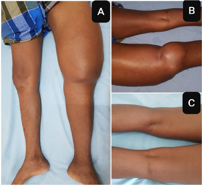
Clinical appearance before and after therapy. (A) Swelling of the left thigh and knee, anterior aspect. (B) Swelling of the left thigh and knee, posterior aspect. (C) Reduction in the swelling of the left thigh and knee, posterior aspect, after therapy.
Laboratory investigations demonstrated elevated inflammatory markers with an ESR of 46 mm in the first hour and CRP of 12 mg/L. Serum uric acid was 9.2 mg/dL. Serum creatinine was mildly elevated at 1.4 mg/dL, with an estimated glomerular filtration rate (eGFR) of 49 mL/min/1.73 m2, consistent with stable chronic kidney disease (Table 1).
| Parameter | At Presentation | 3-month Follow-up | Reference Range |
|---|---|---|---|
| Hemoglobin (g/dL) | 12 | 12.2 | 13-17 |
| Total leukocyte count (cells/mm3) | 4,600 | 6,400 | 4,000-11,000 |
| Platelet count (cells/mm3) | 2.75 lakh | 2.25 lakh | 1.5-4.0 lakh |
| Mean corpuscular volume (fL) | 80 | 80 | 80-100 |
| Blood urea (mg/dL) | 30 | 34 | 15-40 |
| Serum creatinine (mg/dL) | 1.4 | 1.3 | 0.7-1.3 |
| Serum sodium (mEq/L) | 136 | 137 | 135-145 |
| Serum potassium (mEq/L) | 4.4 | 4.6 | 3.5-5.0 |
| Serum chloride (mEq/L) | 100 | 102 | 98-107 |
| Total bilirubin (mg/dL) | 1.0 | 1.0 | 0.3-1.2 |
| AST/SGOT (IU/L) | 29 | 30 | 10-40 |
| ALT/SGPT (IU/L) | 30 | 12 | 7-56 |
| Alkaline phosphatase (IU/L) | 102 | 120 | 44-147 |
| Serum albumin (g/dL) | 4.2 | 4.0 | 3.5-5.0 |
| ESR (mm in first hour) | 46 | 15 | 0-15 |
| CRP (mg/L) | 12 | <6 | <6 |
| Peripheral blood smear | Normal | - | - |
| Serum uric acid (mg/dL) | 9.2 | 7.0 | 3.5-7.2 |
| Serum calcium (mg/dL) | 9.4 | 9.2 | 8.5-10.5 |
| Serum phosphate (mg/dL) | 4.1 | 3.0 | 2.5-4.5 |
| HbA1c (%) / fasting glucose (mg/dL) | 5.8 / 92 | - | <5.7 / 70-99 |
2.1. Diagnostic Methodology
Serum uric acid was estimated using an enzymatic colorimetric microplate assay kit. Under strict aseptic precautions, ultrasound-guided aspiration and drainage of the cyst was performed by an experienced clinician using a 21-gauge needle. Approximately 500 mL of straw-coloured fluid was aspirated for both diagnostic evaluation and therapeutic decompression. No immediate or delayed procedural complications occurred, and repeat aspiration was not required.
Magnetic resonance imaging was performed using a 1.5-T scanner with multiplanar imaging, including coronal T2-weighted Short Tau Inversion Recovery (STIR) sequences together with contrast-enhanced images. Images were interpreted by an experienced consultant radiologist (MD Radiodiagnosis with 10 years of expertise). MRI demonstrated a large multiloculated fluid-filled Baker's cyst communicating with the knee joint, extending superiorly within the intermuscular plane between the quadriceps muscles for approximately 25 cm, measured from the knee joint line to the proximal extent of the cyst on coronal T2-STIR images, displacing the quadriceps tendon and quadriceps muscle anteriorly (Fig. 2A).
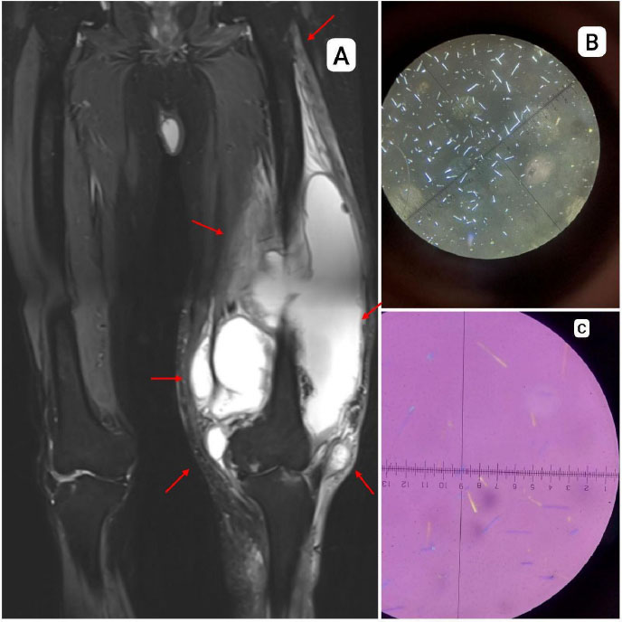
Imaging and synovial-fluid microscopy. (A) MRI of the thigh showing a large multiloculated fluid-filled cyst communicating with the knee joint. (B) Needle-shaped monosodium urate crystals in synovial fluid viewed by light microscopy. (C) Needle-shaped, negatively birefringent monosodium urate crystals in synovial fluid viewed by polarized light microscopy.
Synovial fluid was subjected to cell count, Gram stain, bacterial culture, cytological examination, and polarized light microscopy. Cytological examination revealed no malignant cells. Gram stain, bacterial culture, and cartridge-based nucleic acid amplification test (CB-NAAT) for Mycobacterium tuberculosis were all negative. Polarized microscopy was performed using an Olympus BH-2 BHTP polarized light microscope. Light microscopy demonstrated numerous needle-shaped crystals (Fig. 2B). Polarized light microscopy confirmed that these were negatively birefringent monosodium urate crystals, establishing the diagnosis of gout (Fig. 2C). The complete synovial fluid analysis is summarized in Table 2.
2.2. Differential Diagnosis
The differential diagnosis included soft tissue sarcoma, intramuscular abscess, hematoma, deep vein thrombosis, popliteal artery aneurysm, and other synovial or ganglion cystic lesions [4, 9]. Soft tissue sarcoma was considered because of the large size and unusual proximal extension of the lesion. However, MRI demonstrated a well-defined multiloculated cystic lesion communicating with the knee joint without any solid enhancing component. Furthermore, cytological examination of the aspirated fluid showed no malignant cells, making malignancy unlikely. Intramuscular abscess was excluded because the patient had no fever, leukocytosis, or other systemic features of infection. Gram stain, bacterial culture, and CB-NAAT were all negative. Hematoma was considered unlikely because there was no history of trauma, anticoagulant use, or MRI evidence of blood products at different stages. Deep vein thrombosis was excluded based on the absence of pain, lack of Doppler evidence of venous thrombosis, and characteristic MRI findings demonstrating a cyst communicating with the knee joint. Popliteal artery aneurysm was excluded by the absence of internal vascular flow or arterial continuity on Doppler examination and MRI. Other synovial or ganglion cystic lesions were considered unlikely because the lesion showed the characteristic communication between the knee joint and the gastrocnemius-semimembranosus bursa.
The demonstration of negatively birefringent monosodium urate crystals established the diagnosis of a Baker's cyst secondary to gout.
| Parameter | Result |
|---|---|
| Appearance | Turbid |
| Color | Yellow |
| Total cell count (cells/mm3) | 30 |
| Neutrophils | 90% |
| Monosodium urate crystals | Present |
| Birefringence | Negative |
| Gram stain | Negative |
| Culture and sensitivity | No growth |
| CB-NAAT | Negative |
| Cytology for malignant cells | Negative |
2.3. Treatment
The patient underwent ultrasound-guided therapeutic aspiration of the cyst. Oral prednisolone was initiated at 0.5 mg/kg/day (25 mg/day) for one week, followed by tapering by 5 mg every week and stopped over two months. Febuxostat was started at 40 mg once daily and increased to 80 mg once daily after two weeks, and continued, considering the patient's stable renal function and clinical response. The patient was also advised regarding adherence to urate-lowering therapy and lifestyle modifications.
2.4. Follow-up
At three-month follow-up, the swelling had markedly reduced (Fig. 1C), inflammatory markers had normalized
(ESR 15 mm in the first hour, CRP <6 mg/L, within the laboratory reference range), and serum uric acid had decreased to 7.0 mg/dL (Table 1). The patient remained symptom-free with complete functional recovery.
A follow-up MRI of the left knee and thigh was not performed, as the patient demonstrated marked clinical improvement with significant reduction in swelling and normalization of laboratory parameters. The chronological course of the patient's presentation, investigations, treatment, and three-month follow-up is summarized in Fig. (3).
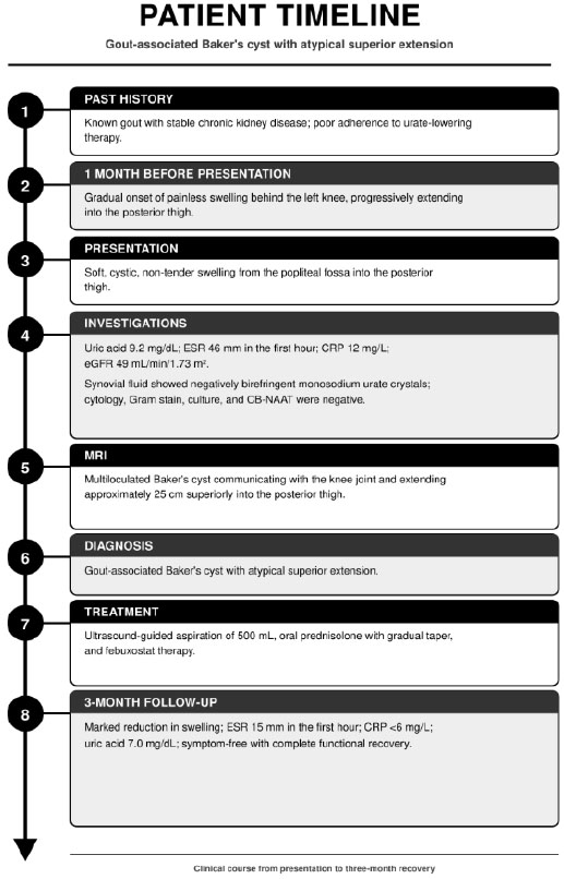
Patient timeline showing the clinical course from presentation to three-month follow-up.
3. DISCUSSION
Baker's cysts develop due to distension of the gastrocnemius-semimembranosus bursa through a valvular communication with the knee joint in the setting of chronic synovitis and increased intra-articular pressure. They are commonly associated with inflammatory arthritis, osteoarthritis, meniscal tears, and other intra-articular pathologies. Inferior extension into the calf is the usual pattern, whereas superior extension into the posterior thigh is rare [1-4, 7].
In gout, deposition of monosodium urate crystals can sustain synovial inflammation, increasing synovial-fluid production and intra-articular pressure. When a communication exists between the knee joint and the gastrocnemius-semimembranosus bursa, a one-way valvular mechanism permits fluid to enter the bursa while limiting its return to the joint. Continued fluid production and pressure progressively enlarge the cyst; the expanding cyst may then dissect along the path of least resistance through intermuscular planes, explaining the marked superior extension observed in this patient [1, 4].
Although Baker's cysts have been reported in association with crystal arthropathies, extensive superior extension into the thigh remains exceedingly rare. Similar atypical presentations have been described in isolated case reports involving intramuscular, dissecting, and giant Baker's cysts, highlighting the potential for these lesions to mimic soft tissue sarcomas, abscesses, or hematomas [7, 8, 10, 11, 13].
MRI remains the imaging modality of choice for atypical Baker's cysts because it accurately demonstrates cyst morphology, communication with the knee joint, extent of anatomical spread, and relationship to adjacent neurovascular and muscular structures. In the present case, MRI clearly demonstrated a multiloculated cyst communicating with the knee joint and extending approximately 25 cm superiorly within the intermuscular plane, thereby excluding important differential diagnoses [1, 4, 7-9].
Although MRI strongly suggested a benign cystic lesion, additional investigations were performed to improve diagnostic confidence. Cytological examination of the aspirated fluid demonstrated no malignant cells, while Gram stain, bacterial culture, and CB-NAAT were negative. The demonstration of negatively birefringent monosodium urate crystals established gout as the underlying etiology [12].
Management of Baker's cysts should primarily target the underlying intra-articular inflammatory process rather than the cyst itself. Our patient underwent ultrasound-guided aspiration for symptomatic decompression followed by systemic corticosteroid therapy and optimization of urate-lowering treatment. Clinical improvement was accompanied by normalization of inflammatory markers and reduction in serum uric acid levels, emphasizing that effective treatment of gout can result in satisfactory resolution without surgical intervention [1, 2, 4, 12, 14].
This case contributes to the limited literature describing extensive superior extension of Baker's cysts associated with gout and reinforces the importance of considering this diagnosis in patients presenting with an unusual posterior thigh mass [7, 8, 10, 11, 13].
CONCLUSION
We report a rare case of a gout-associated Baker's cyst with atypical superior extension into the posterior thigh. MRI accurately demonstrated the lesion and its communication with the knee joint, while synovial fluid crystal analysis, cytology, and microbiological investigations established the diagnosis and excluded important differential diagnoses. Conservative management with ultrasound-guided aspiration, corticosteroid therapy, and optimized urate-lowering treatment resulted in an excellent clinical outcome.
LIMITATIONS
This report describes a single patient and therefore cannot determine the frequency or causal relationship between gout and extensive superior extension of Baker's cysts. Follow-up MRI was not performed because the patient demonstrated marked clinical and biochemical improvement following conservative management. Histopathological confirmation was not pursued because MRI findings, synovial fluid crystal analysis, negative cytology, and negative microbiological investigations were considered sufficient to establish the diagnosis.
LEARNING POINTS
- Baker's cysts usually extend inferiorly into the calf; superior extension into the posterior thigh is rare.
- Gout-related chronic synovitis can predispose to the development of atypically large Baker's cysts.
- MRI is the preferred imaging modality for defining the extent of the lesion and excluding important differential diagnoses.
- Synovial fluid crystal analysis, together with cytology and microbiological investigations when indicated, improves diagnostic certainty.
- Conservative treatment directed at the underlying inflammatory disorder, combined with therapeutic aspiration when required, can achieve excellent outcomes without surgical intervention.
PATIENT PERSPECTIVE
I noticed a painless swelling behind my knee. It slowly got bigger and extended into my thigh. I was worried that it might be something serious.
The doctors did some tests and told me that the swelling was related to my gout. They explained that it was not a tumor or an infection. This helped me and my family feel less worried.
The doctors drained the swelling and gave me medicines for gout. Over the next few weeks, the swelling got smaller, and I felt better. I now understand why I need to take my gout medicines regularly and come back for check-ups.
AUTHORS’ CONTRIBUTIONS
The authors confirm their contributions to the paper as follows: All authors attest that they meet the current International Committee of Medical Journal Editors (ICMJE) criteria for authorship. All authors have read and agreed to the published version of the manuscript.
LIST OF ABBREVIATIONS
| CB-NAAT | = Cartridge-Based Nucleic Acid Amplification Test |
| CRP | = C-Reactive Protein |
| ESR | = Erythrocyte Sedimentation Rate |
| MRI | = Magnetic Resonance Imaging |
| STIR | = Short Tau Inversion Recovery |
CONSENT FOR PUBLICATION
Written informed consent was obtained from the patient for the publication of clinical details and images.
AVAILABILITY OF DATA AND MATERIALS
All data generated or analyzed during this study are included in this published article.
CONFLICT OF INTEREST
The authors have no financial conflicts to disclose. All authors have reviewed and approved the manuscript, and all authors fulfill the criteria for authorship.
ACKNOWLEDGEMENTS
The authors acknowledge all colleagues who assisted or collaborated in the study.