All published articles of this journal are available on ScienceDirect.

Biopsy-Proven Lupus Nephritis in a Patient Living with HIV: Autoimmunity in Immunodeficiency- A Case Report

Abstract
Background
The coexistence of Systemic Lupus Erythematosus (SLE) and Human Immunodeficiency Virus (HIV) infection represents a rare and complex clinical entity due to opposing immunological mechanisms. While HIV leads to immunodeficiency through CD4+ T-cell depletion, SLE is characterized by immune hyperactivity and autoantibody production.
Case Presentation
We report a 50-year-old female, a known Person Living with HIV (PLHIV) on Highly Active Antiretroviral Therapy (HAART), who presented with polyarthritis, alopecia, and significant weight loss. Laboratory evaluation revealed pancytopenia, markedly elevated anti-dsDNA antibodies, hypocomplementemia, and significant proteinuria. HIV disease was well controlled, with a CD4 count of 600 cells/mm3 and a low viral load of 120 copies/mL at presentation. Renal biopsy demonstrated focal mesangial and endocapillary hypercellularity, while immunofluorescence showed granular deposition of IgG, C3, and C1q, confirming Class 3 (ISN/RPN) lupus nephritis. The patient was treated with corticosteroids, hydroxychloroquine, and mycophenolate mofetil, along with cotrimoxazole prophylaxis and continuation of HAART. On follow-up after 6 months, she achieved complete renal remission with stable HIV disease status and is currently maintained on low-dose corticosteroids, mycophenolate mofetil, hydroxychloroquine, and HAART.
Conclusion
This case highlights the diagnostic and therapeutic challenges of autoimmune disease in an immunocompromised host and underscores that effective viral suppression with preserved immune status permits safe and effective use of standard immunosuppressive therapy.
1. INTRODUCTION
The coexistence of Systemic Lupus Erythematosus (SLE) and Human Immunodeficiency Virus (HIV) infection represents a paradox in clinical immunology. SLE is characterized by immune hyperactivity, autoantibody production, and immune complex deposition, whereas HIV infection leads to progressive immunodeficiency through CD4+ T-cell depletion. Historically, autoimmune diseases were thought to be suppressed in HIV-infected individuals. However, the introduction of Highly Active Antiretroviral Therapy (HAART) has altered the disease spectrum, with increasing reports of autoimmune manifestations including SLE [1-4].
Several mechanisms have been proposed to explain this phenomenon, including Immune Reconstitution Inflammatory Syndrome (IRIS), molecular mimicry, chronic immune activation, and dysregulated immune responses despite virologic suppression [2-6]. Persistent antigenic stimulation and impaired regulatory T-cell function in HIV infection may facilitate autoreactive B-cell expansion and subsequent autoimmune disease development [3-6].
Renal involvement in HIV is heterogeneous and includes HIV-Associated Nephropathy (HIVAN) and HIV-associated immune complex kidney disease (HIVICK), both of which may mimic lupus nephritis clinically and histologically [7-11]. Therefore, distinguishing true lupus nephritis from HIV-associated renal disease is crucial for appropriate management because treatment strategies and prognosis differ substantially.
2. CASE PRESENTATION
A 50-year-old female, a known person living with HIV for 8 years on HAART (tenofovir 300mg, lamivudine 300mg, dolutegravir 50 mg), presented with polyarthritis, alopecia, and significant weight loss of 10 kg over 6 months. She had a past history of pulmonary tuberculosis 6 years back and was treated for the same. She had no other known comorbid conditions.
On examination, vitals were stable. She was thin-built and had malar rash and non-scarring alopecia (Figure 1A, B). She was afebrile, had pallor, and generalized lymphadenopathy. Examination of Cardiovascular, respiratory, and central nervous system were normal. Abdomen examination revealed mild hepatosplenomegaly. Musculoskeletal examination showed polyarthritis involving small and large joints of both upper and lower limbs.
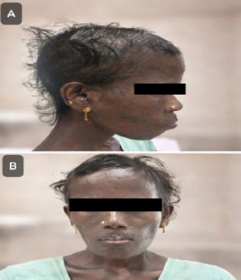
(A) Anterior view showing malar rash, alopecia. (B) Lateral view showing malar rash, alopecia.
Laboratory evaluation revealed pancytopenia. Random blood sugars, renal and liver function tests were normal. Urine analysis revealed significant proteinuria. ANA profile showed strongly positive anti-dsDNA antibodies, along with anti-nucleosome, antihistone, and anti-Smith antibodies. Complement levels were reduced. HIV testing confirmed HIV-1 positivity with a CD4 count of 600 cells/mm3 and low viral load. (Tables 1, 2). Ultrasonogram of the abdomen showed mild hepatosplenomegaly, with no significant lymphadenopathy and normal-sized kidneys.
| Parameter | D(0) | D(90) | D(120) |
|---|---|---|---|
| CBC | |||
| Hemoglobin (g/dL) | 8.7 | 10.6 | 12.1 |
| WBC (/µL) | 2870 | 4000 | 7100 |
| Neutrophil/Lymphocyte (%) | 53/36 | 60/32 | 62/32 |
| Platelets (×105/µL) | 1.47 | 1.64 | 2.31 |
| Hematocrit (%) | 40 | 40 | 39 |
| MCV (fL) | 82 | 84 | 80 |
| MCH (pg) | 33 | 31 | 30 |
| MCHC (g/dL) | 32 | 31 | 30 |
| Acute Phase Reactants | |||
| ESR (mm/hr) | 28 | 30 | 26 |
| CRP (mg/L) | Negative | Negative | Negative |
| RBS (mg/dL) | 60 | 84 | 92 |
| RFT and LFT | |||
| Urea (mg/dL) | 40 | 42 | 36 |
| Creatinine (mg/dL) | 0.6 | 0.7 | 0.6 |
| Sodium (mEq/L) | 140 | 142 | 136 |
| Potassium (mEq/L) | 3.7 | 3.6 | 3.8 |
| AST (U/L) | 18 | 14 | 18 |
| ALT (U/L) | 13 | 16 | 16 |
| Total Bilirubin (mg/dL) | 0.3 | 0.4 | 0.3 |
| Direct Bilirubin (mg/dL) | 0.1 | 0.1 | 0.1 |
| Total Protein / Albumin (g/dL) | 5.8/2.8 | 6.6/4.0 | 6.5/4.1 |
| ALP (U/L) | 64 | 60 | 60 |
| Urine Analysis | |||
| Urine Albumin | 2+ | 2+ | Nil |
| Spot PCR | 1.8 | 1.1 | 0.3 |
| 24-hour Urine Protein (mg/day) | 1600 | 400 | 230 |
| SLE Disease Activity | D (0) | D(120) | |
|---|---|---|---|
| Anti-dsDNA (IU/mL) | 477 | - | 40 |
| C3 (mg/dL) | 0.29 | - | 1.1 |
| C4 (mg/dL) | 0.09 | - | 0.2 |
| HIV Disease Monitoring | - | - | - |
| CD4 Count (cells/mm3) | 600 | - | 580 |
| HIV Viral Load (copies/mL) | 120 | - | 130 |
Renal biopsy demonstrated focal mesangial and endocapillary hypercellularity on light microscopy (Figure 2), while immunofluorescence revealed granular deposition of IgG, C3, and C1q along the capillary loops and mesangium (Figure 3A-C) consistent with lupus nephritis.
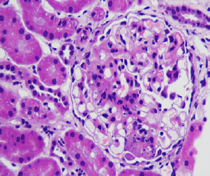
Light microscopy showing focal mesangial hypercellularity and endocapillary hypercellularity suggestive of focal glomerular nephritis.
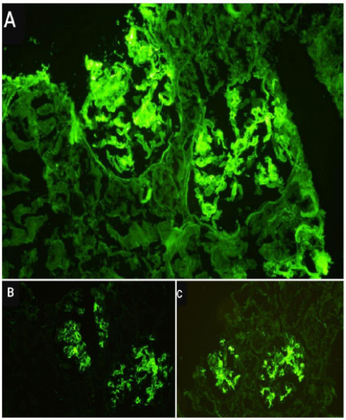
Immunofluorescence staining showing granular positivity on capillary loops and mesangium for IgG Fig 3 (A); C3 Fig 3 (B); C1q Fig 3 (C).
Based on clinical, serological, and histopathological findings, a diagnosis of systemic lupus erythematosus with lupus nephritis in the background of HIV infection was established.
The patient was initiated and treated with corticosteroids (intravenous pulse steroids 500mg once a day for 3 days followed by prednisolone 50mg/day, tapering dose of oral steroids by 5mg every 2 weeks, reached 5 mg/day by six months), hydroxychloroquine 200mg once a day, mycophenolate mofetil 1gm/day in two divided doses along with antiproteinuric therapy (enalapril 10mg/day in two divided doses) and cotrimoxazole(80/400 mg once a day) prophylaxis for SLE and lupus nephritis. Close monitoring of CD4 counts and viral load was performed, and she was continued with HAART.
She showed significant clinical improvement with resolution of symptoms and reduction in proteinuria, achieving remission of lupus nephritis. Serial monitoring showed progressive improvement in disease activity: anti-dsDNA titres fell from 477 IU/mL to 40 IU/mL, C3 and C4 levels normalized, haemoglobin improved from 8.7 to 12.1 g/dL, and 24-hour urinary protein fell from 1600 mg/day to 230 mg/day between day 0 and day 120 of treatment (Tables 1, 2). There was significant resolution of arthralgia by 2 months, weight gain by 3 months, and visible hair regrowth by 4 months of therapy. Her HIV disease was also under control as evidenced by normal CD4 count and low viral load on follow-up. She is currently on mycophenolate mofetil (1 gm/day), hydroxychloroquine (200mg/day), corticosteroids (prednisolone 5mg/day), HAART regimen (Tenofovir 300mg, Lamivudine 300mg, Dolutegravir 50mg) and cotrimoxazole prophylaxis. She is under regular follow-up and being monitored for both SLE disease activity and HIV disease activity. At the time of this report, she continues on this regimen with sustained renal remission, preserved renal function, and stable, virologically suppressed HIV disease, with no lupus flare or opportunistic infection since the initial six-month assessment, reflecting her current clinical status.
3. DISCUSSION
The coexistence of Systemic Lupus Erythematosus (SLE) and Human Immunodeficiency Virus (HIV) infection represents a unique immunological paradox. While HIV infection leads to immune suppression through CD4+ T-cell depletion, it can paradoxically promote autoimmunity through mechanisms such as polyclonal B-cell activation, molecular mimicry, chronic immune activation, and dysregulated cytokine production despite virologic suppression [3-6]. Additionally, immune reconstitution following Highly Active Antiretroviral Therapy (HAART) has been implicated in triggering autoimmune diseases [2-4].
Recent evidence suggests that chronic immune activation in HIV, even in virologically suppressed patients, plays a pivotal role in autoimmunity. Persistent antigenic stimulation leads to expansion of autoreactive B-cell clones and impaired regulatory T-cell (Treg) function, creating a permissive environment for autoimmune diseases such as SLE [3-6]. Another important mechanism is immune reconstitution Inflammatory Syndrome (IRIS), where rapid restoration of CD4+ T-cell function following HAART triggers exaggerated immune responses against self-antigens. Cases of SLE emerging after HAART initiation support this phenomenon [2-4].
Renal involvement in HIV-infected individuals is multifactorial. HIV-associated Immune Complex Kidney disease (HIVICK) can closely resemble lupus nephritis both clinically and histologically [7-11]. However, certain features help differentiate the two. True lupus nephritis is characterized by high-titre anti-dsDNA antibodies, hypocomplementemia, and a classical “full-house” immunofluorescence pattern [7]. Our patient demonstrated all these features, confirming the diagnosis. The distinction between HIVICK and lupus nephritis is clinically critical. HIVICK typically demonstrates dominant IgA or mixed immune deposits with less consistent complement consumption, whereas lupus nephritis shows classical “full-house” staining with IgG, IgA, IgM, C3, and C1q deposition [7-10]. Moreover, lupus nephritis is strongly associated with systemic features such as cytopenias, arthritis, and serologic autoimmunity [1, 2, 7, 9, 10].
Several case reports and observational studies have documented the coexistence of SLE and HIV, although the condition remains rare [1-4, 12]. Patients with HIV-associated SLE often present after immune restoration with relatively preserved CD4 counts, supporting the concept that immune reconstitution permits emergence of autoimmunity [1-4]. In contrast to previous reports describing lower anti-dsDNA positivity in HIV-associated SLE, our patient demonstrated markedly elevated anti-dsDNA titres and hypocomplementemia, suggesting a more classical and immunologically active SLE phenotype [1-4]. Quantitative data from dedicated cohorts underscore this rarity: in a 25-year single-centre US HIV-rheumatology clinic cohort, only 22 of 1,604 patients (1.4%) developed concomitant SLE, with African-American women disproportionately affected (81.8% vs. 55.3% of the general clinic population, p=0.02) [13]. Similarly, a French multicentre case-control study spanning 1975–2020 identified only 13 patients with SLE-HIV overlap, in whom renal involvement (54% vs. 16%, p=0.006) and autoimmune cytopenias (31% vs. 8%, p=0.04) were significantly more frequent than in matched HIV-negative SLE controls [14]. In a large Spanish cohort of 5,665 people living with HIV, the overall prevalence of autoimmune disease was 5.3% (95% CI 4.7–5.9) [3]. These figures confirm that SLE-HIV overlap, while genuinely uncommon, is associated with a distinct and clinically more severe renal phenotype, consistent with our patient's presentation.
Broader immunologic studies have further demonstrated that HIV infection is characterized by sustained immune activation and dysregulation despite effective viral suppression [5, 6]. Haas et al. demonstrated that HIV-associated immune complex glomerulonephritis may exhibit “lupus-like” features but typically lacks high-titre anti-dsDNA antibodies and hypocomplementemia [7]. In contrast, our patient demonstrated high anti-dsDNA titres, low complement levels, and classical “full-house” immunofluorescence findings, confirming true lupus nephritis. This distinction is clinically important because HIV-associated kidney disease may additionally be influenced by antiretroviral therapy-related nephrotoxicity [11], whereas lupus nephritis requires targeted immunosuppressive therapy.
An important consideration is whether persistent HIV infection itself, or the antiretroviral agents used to control it, alter the histologic appearance of the kidney in a way that could confound the diagnosis of lupus nephritis. Classic HIV-Associated Nephropathy (HIVAN) is a podocytopathy characterized by collapsing focal segmental glomerulosclerosis with microcystic tubular dilatation and interstitial inflammation, occurring predominantly in patients with uncontrolled viraemia and advanced immunosuppression, and it is not typically associated with immune-complex deposition [7, 8]. Tenofovir, part of our patient's HAART regimen, is well recognized to cause a distinct pattern of proximal tubular injury and mitochondrial toxicity (acute tubular necrosis, Fanconi-like tubulopathy) rather than glomerular disease, and this tubulointerstitial pattern is readily distinguished histologically from the glomerular, immune-complex-mediated injury of lupus nephritis [11, 15]. In our patient, HIV viraemia was well suppressed (120 copies/mL) at the time of biopsy, and renal histology showed a proliferative, immune-complex glomerulonephritis with full-house immunofluorescence (IgG, C3, C1q) rather than the collapsing glomerulopathy or tubular changes attributable to HIVAN or antiretroviral nephrotoxicity. This argues against a major contribution of persistent HIV replication or HAART to the observed histologic alterations and supports a primarily autoimmune, lupus-driven process. Nonetheless, because tenofovir-associated tubulopathy and lupus nephritis can coexist, renal function and urinary findings should continue to be monitored longitudinally to detect any superimposed drug-related tubular injury.
Beyond acquired immunodeficiency states such as HIV infection, several congenital (primary) immunodeficiencies are well recognized to predispose to SLE, most notably homozygous deficiencies of the early classical complement pathway components C1q, C1r/s, C2, and C4, as well as common variable immunodeficiency (CVID) [16]. C1q deficiency in particular confers a very high risk (over 90%) of developing an SLE-like illness, typically with early-onset (childhood or adolescent), severe multi-organ disease including nephritis [16]. The pathogenic mechanism in these congenital disorders centres on impaired opsonization and clearance of apoptotic cellular debris and immune complexes, which promotes chronic exposure of nuclear autoantigens to the immune system and breakdown of self-tolerance from birth [16]. This differs fundamentally from the acquired immunodeficiency of HIV infection, in which autoimmunity typically emerges later in the disease course, often during immune reconstitution on HAART, and is driven by chronic immune activation, polyclonal B-cell hyperactivity, and dysregulated T-cell help rather than a primary defect in immune-complex clearance [2-6]. Our patient, who developed lupus nephritis after several years of virologically suppressed HIV infection with a preserved CD4 count, exemplifies this acquired, immune-reconstitution-associated pathway rather than a congenital clearance defect; she had no history suggestive of early-onset or recurrent severe infections that might point to an underlying primary immunodeficiency. Nonetheless, the shared endpoint in both congenital and acquired immunodeficiency states, a permissive environment for loss of self-tolerance, reinforces the broader concept that immunodeficiency, whatever its origin, can paradoxically facilitate rather than prevent autoimmune disease.
Management of lupus nephritis in patients with HIV infection poses a therapeutic challenge. Immunosuppressive therapy increases the risk of opportunistic infections and can potentially worsen viral replication. However, studies suggest that patients with stable HIV disease and adequate CD4 counts can safely receive standard immunosuppressive therapy with careful monitoring [1-4]. Balancing immunosuppressive therapy with ongoing antiretroviral treatment remains essential.
From a therapeutic standpoint, Mycophenolate Mofetil (MMF) is particularly advantageous because of its selective inhibition of lymphocyte proliferation and relatively favorable safety profile compared with cyclophosphamide [17-19]. Hydroxychloroquine provides additional benefit through immunomodulatory and potential antiviral effects, including inhibition of Toll-like receptor signaling [17-19].
Drug–drug interactions must also be carefully considered, especially between HAART and immunosuppressive agents. Protease inhibitors may alter corticosteroid metabolism, increasing the risk of iatrogenic Cushing syndrome and adrenal suppression, necessitating vigilant monitoring [11, 17-19]. Long-term prognosis in such patients depends on maintaining an optimal balance between immune suppression and infection control. Available evidence suggests that patients with well-controlled HIV infection, preserved CD4 counts, and suppressed viral load can achieve outcomes comparable to HIV-negative lupus nephritis when treated appropriately [17-19].
Finally, this case reinforces the evolving paradigm that HIV infection is not merely a state of immunodeficiency but rather one of immune dysregulation, capable of manifesting both opportunistic infections and autoimmune diseases simultaneously [3-6].
4. STUDY LIMITATIONS
As a single case report, this study does not permit statistical inference or generalisation beyond the individual patient described, and no comparison group or hypothesis testing was applicable given the descriptive design. Disease activity was assessed using serial serological (anti-dsDNA, C3, C4) and renal (proteinuria, urine albumin) parameters (Tables 1 and 2) rather than a formal validated composite index such as SLEDAI-2K or BILAG, and granular timelines for individual clinical manifestations (e.g., alopecia regrowth, weight regain) were not systematically recorded. No repeat renal biopsy was performed to histologically confirm remission, which was inferred from clinical and laboratory parameters. Formal complement genetic testing or a dedicated primary immunodeficiency work-up was not undertaken to exclude an underlying congenital immune defect, although the patient's history did not suggest one. Finally, the follow-up duration, while demonstrating sustained remission, remains relatively short for assessing long-term outcomes such as lupus nephritis relapse or HIV virological rebound.
CONCLUSION
Lupus nephritis in patients living with HIV is rare and presents significant diagnostic and therapeutic challenges. Differentiation from HIV-associated renal disease is critical and relies on serological and histopathological confirmation. With appropriate selection and monitoring, standard immunosuppressive therapy can be safely administered, leading to good clinical outcomes.
PATIENT’S PERSPECTIVE
“I have been living with HIV for many years and was doing well on regular treatment. When I started developing joint pains, hair loss, and weight loss, I was worried that my HIV was worsening. After several tests, I was told that I had lupus affecting my kidneys, which was unexpected and frightening for me.
Initially, I was anxious about taking additional medications, especially immunosuppressive drugs, because of my existing HIV condition. However, my doctors explained the condition clearly and reassured me about close monitoring. With treatment, I gradually started feeling better; my joint pains improved, and my overall health recovered.
Now, I feel more confident continuing both my HIV treatment and the medications for lupus. Regular follow-up and support from my healthcare team have helped me manage both conditions. I hope my experience helps others understand that even with HIV, such conditions can be treated successfully.”
AUTHORS’ CONTRIBUTIONS
The authors confirm contribution to the paper as follows: A.K.: Study conception and design; D.K.: Data collection. A.R.: Analysis and interpretation of results. A.K.: Draft manuscript. All authors reviewed the results and approved the final version of the manuscript.
LIST OF ABBREVIATIONS
| SLE | = Systemic Lupus Erythematosus |
| HIV | = Human Immunodeficiency Virus |
| PLHIV | = Person Living with Human Immunodeficiency Virus |
| HAART | = Highly Active Antiretroviral Therapy |
| IRIS | = Immune Reconstitution Inflammatory Syndrome |
| ANA | = Antinuclear Antibody |
| Anti-dsDNA | = Anti-double-stranded DNA Antibody |
| C3 | = Complement Component 3 |
| C4 | = Complement Component 4 |
| CBC | = Complete Blood Count |
| WBC | = White Blood Cell Count |
| MCV | = Mean Corpuscular Volume |
| MCH | = Mean Corpuscular Hemoglobin |
| MCHC | = Mean Corpuscular Hemoglobin Concentration |
| ESR | = Erythrocyte Sedimentation Rate |
| CRP | = C-reactive Protein |
| RFT | = Renal Function Test |
| LFT | = Liver Function Test |
| RBS | = Random Blood Sugar |
| PCR | = Protein-to-Creatinine Ratio |
| ALP | = Alkaline Phosphatase |
| AST | = Aspartate Aminotransferase |
| ALT | = Alanine Aminotransferase |
| IF | = Immunofluorescence |
| ISN/RPS | = International Society of Nephrology/Renal Pathology Society |
| HIVAN | = HIV-associated Nephropathy |
| HIVICK | = HIV-associated Immune Complex Kidney Disease |
| CVID | = Common Variable Immunodeficiency |
HUMAN AND ANIMAL RIGHTS
All procedures involving human participants were conducted in accordance with the ethical standards of the committee responsible for human experimentation (institutional and national), and with the Helsinki Declaration of 1975, as revised in 2013.
CONSENT FOR PUBLICATION
Written informed consent for publication of this case report, including the clinical photographs (Figure 1), histopathology images, and associated clinical data, was obtained from the patient.
AVAILABILITY OF DATA AND MATERIAL
All data generated or analyzed during this study are included in this published article.
ACKNOWLEDGEMENTS
Declared none.