All published articles of this journal are available on ScienceDirect.

Diagnostic Challenges: A Case Series of Sarcoidosis
 iD
Arul Raja Murugan PS2
iD
Dhiliprajan Kannan2
iD
Authors Info & Affiliations
Abstract
Introduction
Sarcoidosis is a multisystem granulomatous disease of unknown etiology characterized histologically by non-caseating granulomas. Diagnosis is often by exclusion, requiring the elimination of infectious and foreign-body triggers. While bilateral hilar lymphadenopathy and elevated serum Angiotensin-Converting Enzyme (ACE) levels are suggestive, their diagnostic sensitivity and specificity are limited. Furthermore, clinical management is often guided by anecdotal evidence due to lack of randomized controlled trials, particularly in cases involving extra-pulmonary organs.
Case Presentation
In this case series, we describe three distinct and diagnostically challenging presentations of sarcoidosis involving critical organ systems:
1. Renal involvement: Highlights the diagnostic pathway in a patient presenting with unexplained renal insufficiency.
2. Cardiac involvement: Details the complexities of identifying sarcoidosis as the underlying cause of conduction abnormalities and arrhythmia.
3. Central nervous system (CNS) involvement: Illustrates the “masquerader” nature of neurosarcoidosis and the challenges of obtaining tissue confirmation at neurological sites.
Each case highlights the diagnostic hurdles faced when classic pulmonary findings are absent or subtle, and the reliance on a high index of clinical suspicion alongside multimodal imaging.
Conclusion
These cases underscore the extreme clinical heterogeneity of sarcoidosis. Involvement of the renal, cardiac, and central nervous systems poses a significant diagnostic challenge, as delays in treatment can lead to significant morbidity. This series emphasizes the need for a standardized diagnostic approach and contributes to the growing body of anecdotal evidence that currently guides the management of rare extra-pulmonary manifestations of this “great masquerader.”
1. INTRODUCTION
Sarcoidosis is an autoimmune disease of unknown etiology that can affect any organ in the body. The characteristic involvement is bilateral hilar adenopathy [1]. The histological hallmark of sarcoidosis is the presence of noncaseating granuloma, clusters of macrophages, epithelioid cells, mononuclear cells, and CD4+ T cells, with a few CD8+ T cells, in the peripheral zone [1]. Noncaseating granuloma is not pathognomonic of sarcoidosis. Hence, the diagnosis of sarcoidosis is based on (a) the presence of noncaseating granuloma on histopathologic examination, (b) compatible clinical presentation, and (c) exclusion of other causes of granulomatous inflammation. Diagnosing sarcoidosis of the central nervous system, heart, and eyes is particularly challenging in view of tissue inaccessibility [1]. Herein, we discuss three such difficult-to-diagnose cases of sarcoidosis.
2. CASE PRESENTATION
2.1. Case 1
A middle-aged female with a history of diabetes mellitus for fifteen years presented elsewhere with right ear pain for two days followed by difficulty in chewing on the right side for five days. The sequential events are represented below (Fig. 1).
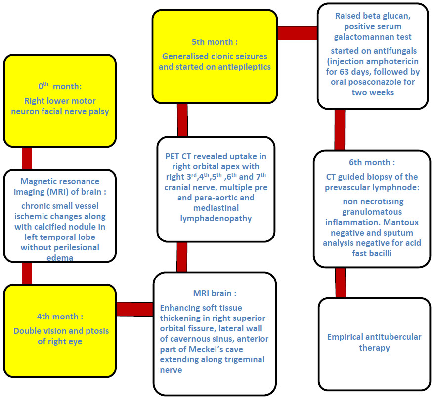
Sequence of the clinical course of Case 1 (neurosarcoidosis).
She was started on antitubercular therapy, which she continued for five months. Repeat MRI revealed persistent abnormal enhancement of the extra-axial soft tissue thickening in the right cavernous sinus, extending up to the orbital apex. The relevant investigations are tabulated (Table 1). Considering the clinicopathological and imaging evidence, a diagnosis of possible neurosarcoidosis was made, and she was started on oral prednisolone followed by steroid-sparing agent methotrexate. Over the period of six months after initiation of treatment, she improved clinically, and she demonstrated paradoxical Mantoux positivity (Fig. 2).
| Variables | Case 1 | Case2 | Case 3 |
|---|---|---|---|
| Type of sarcoidosis | Neuro sarcoidosis | Renal sarcoidosis | Cardiac sarcoidosis |
| Age | 54 | 49 | 23 |
| Sex | female | female | Female |
| Presentation | Multiple cranial nerve palsies | Incidental on screening renal function tests | Exertional dyspnea and palpitation |
| Comorbidities | Diabetes mellitus | Schizophrenia | Acyanotic Congenital heart disease |
| Parotidomegaly | nil | yes | Nil |
| Pulmonary involvement | Mediastinal lymphadenopathy | Right lower lobe bronchial wall thickening, ectasia and tubular bronchiectasis | Nil |
| Mantoux testing | Negative | negative | negative |
| Serum angiotensin-converting enzyme (ACE) (8-65 IU/L) |
8 | 58 | 14.8 |
| Serum calcium (8-10 mg/dl) |
8.6 | 14.4 | 13 |
| Biopsy |
CT-guided biopsy of lymph node: Non-necrotising granulomatous inflammation (Fig. 2) |
Renal biopsy: Ill-defined non-necrotising epithelioid granuloma in two areas of the interstitium with scattered lymphoplasmacytic infiltrates |
Nil |
| Imaging |
MRI Brain: Enhancing soft tissue thickening in right superior orbital fissure, lateral wall of cavernous sinus, anterior part of Meckel’s cave extending along the trigeminal nerve PET CT: Revealed uptake in right orbital apex with right 3rd,4th, 5th, 6th and 7th cranial nerves, multiple pre- and para-aortic and mediastinal lymphadenopathy. |
Ultrasound abdomen and pelvis: Normal-sized kidneys Right kidney - 9*4 cm Left kidney- 8.9*4.2 cm With preserved corticomedullary differentiation. |
CMR: Hypokinesia of the left ventricle with significant thinning of the anteroseptal wall. LGE: (Fig. 4) Sub-epicardial delayed hyper-enhancement in the intraventricular septum indicative of granulomatous involvement. Cardiac PET: focal intense uptake in multiple areas of the left ventricle, decreased uptake in the septum reflecting thinning and fibrosis of myocardium. There was no evidence of extra-myocardial abnormal uptake noted |
| Relevant other investigations |
ANCA (ELISA) - negative for MPO & PR3 Serum IgG4 – within normal limits ANA (IIF): Negative Bronchoalveolar lavage: Negative for infections CD4/CD8 not done |
Normocytic normochromic anaemia Hemoglobin - 8 g/dl Serum urea - 34 mg/dl Serum Creatinine - 1.6 mg/dl ANA(IIF): Negative Sputum gram stain and culture for acid-fast bacilli negative |
ANA(IIF): Negative ECG: biatrial enlargement Chest X-ray: cardiomegaly. Echocardiogram: Dilatation of left-sided chambers of heart along with concentric left ventricular hypertrophy and moderate systolic dysfunction |
| Empirical Antitubercular therapy | Yes For 6 months |
Yes For one month and discontinued in view of intolerance |
nil |
| Treatment | Oral prednisolone at 0.5 mg/kg followed by methotrexate at 15 mg/week | Oral prednisolone at 0.5mg/kg with gradual taper | Oral prednisolone at 1 mg/kg followed by gradual taper over 4-6 months, along with oral methotrexate at 10mg/week along with beta blockers, RAAS inhibitors and diuretics. |
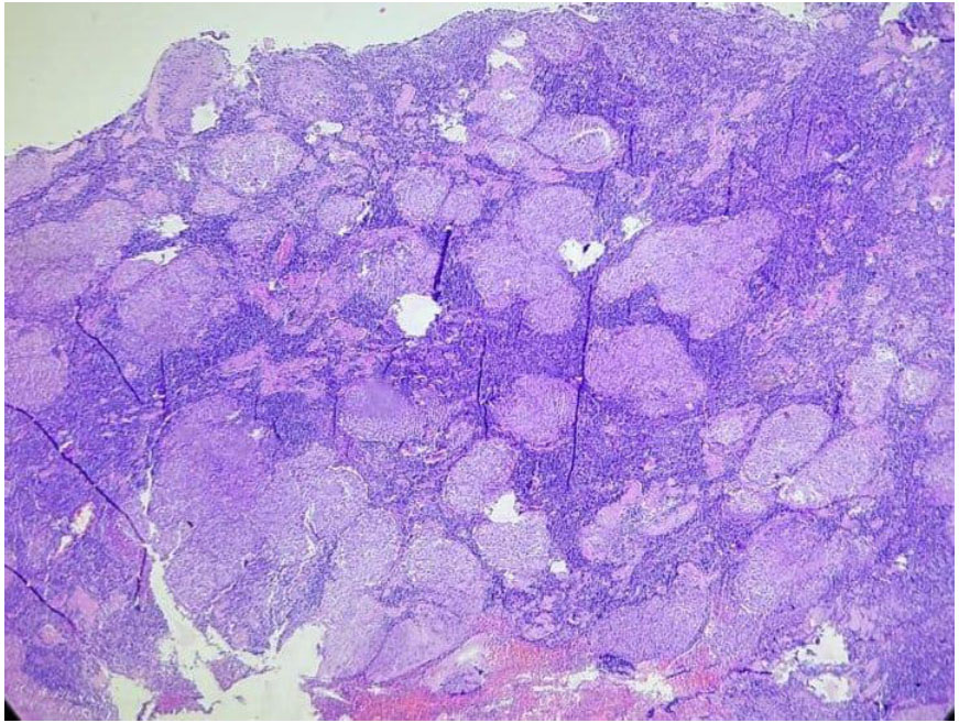
Lymph node biopsy showing non-necrotising granuloma in Case 1 (neurosarcoidosis).
2.2. Case 2
A middle-aged female with a past medical history of schizophrenia for thirteen years, on routine evaluation, was found to have high renal parameters. On evaluation, she was found to have right parotid gland enlargement with normocytic normochromic anemia and increased serum calcium of 14.4 mg/dl. Her further relevant investigations are tabulated in Table 1. Renal biopsy was suggestive of granulomatous interstitial nephritis. She was started on empirical antitubercular therapy, which she discontinued due to intolerance. After eight months, she presented with painful bilateral parotid swelling, and ultrasonogram of the parotid gland revealed heterogeneous echotexture with multiple scattered hypoechoic areas, suggestive of chronic sialadenitis. Fine-needle aspiration cytology of the parotid gland was consistent with chronic sialadenitis. In consideration of the background of biopsy evidence of granulomatous interstitial nephritis with bilateral parotid swelling, possible sarcoidosis was diagnosed and started on oral prednisolone therapy at 0.5 mg/kg. Within 6-8 weeks, the patient responded well and had complete resolution of her parotid swelling (Fig. 3).
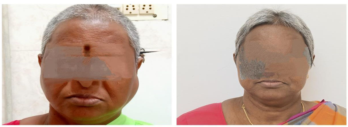
Resolution of parotid swelling following oral corticosteroid therapy in Case 2 (renal sarcoidosis).
2.3. Case 3
A young unmarried woman with a history of small ostium secundum atrial septal defect (OS-ASD) and small perimembranous Ventricular Septal Defect (VSD), both of which reportedly resolved spontaneously by 3 months of age, presented with worsening exertional dyspnoea and palpitations for the past 3 months. Her further relevant investigations are tabulated in Table 1. Cardiac Magnetic Resonance imaging (CMR) revealed thinning of the anteroseptal wall and left ventricular hypokinesia, along with subepicardial Late Gadolinium Enhancement (LGE) involving the interventricular septum consistent with granulomatous infiltration (Fig. 4). Cardiac fluorine-18 fluorodeoxyglucose (FDG)–Positron Emission Tomography (PET) showed moderate to severe reduction in metabolic activity in the interventricular septum, consistent with fibrosis and focal intense FDG uptake in the anterior, lateral, and inferior walls of the left ventricle suggestive of active myocardial inflammation. Coronary angiography revealed normal coronary arteries, thereby ruling out an ischaemic aetiology. A diagnosis of cardiac sarcoidosis was established based on the Japanese Circulation Society (JCS) 2016 criteria. She was treated with oral prednisolone, followed by methotrexate, along with Renin–Angiotensin–Aldosterone System (RAAS) inhibitors, diuretics and beta-blockers. Although she remains clinically stable on follow-up, she is currently being evaluated for cardiac transplantation in view of advanced cardiac involvement.
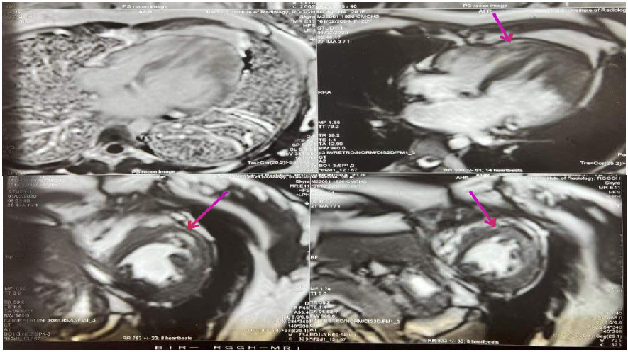
Cardiac magnetic resonance imaging of Case 3 (cardiac sarcoidosis), showing sub-epicardial Late Gadolinium Enhancement (LGE) in the interventricular septum.
3. DISCUSSION
The American Thoracic Society (ATS), European Respiratory Society (ERS), and World Association of Sarcoidosis and Other Granulomatous Disorders (WASOG) define sarcoidosis as a multisystem disorder of unknown aetiology, diagnosed on the basis of clinicoradiological findings in the presence of histological evidence of noncaseating epithelioid granuloma, when other causes of such granuloma have been excluded [2]. However, the 2025 WASOG Task Force and the 2024 American Heart Association (AHA) Scientific Statement recognize varying levels of diagnostic certainty in cases where tissue confirmation is difficult to obtain, such as in cardiac or central nervous system sarcoidosis [3, 4]. Hence, histological demonstration of non-caseating granulomas plays an important role in establishing the diagnosis. Other causes of granulomatous inflammation, including tuberculosis, fungal infections, silicosis and malignancy, must be excluded before establishing a diagnosis of sarcoidosis. Histological confirmation may be difficult in cardiac and neuro-sarcoidosis, particularly when the disease is restricted to a single organ or organ system [2].
Neurosarcoidosis may affect any anatomic structure and is associated with diverse clinical features. Granulomas at the base of the brain parenchyma, with a perivascular distribution, are considered specific for neurosarcoidosis [2]. As meningeal inflammation becomes chronic, progressive collagen deposition by fibroblasts may result in pachymeningeal thickening and subsequent dense fibrosis. However, leptomeningeal involvement is more common than pachymeningeal involvement in neurosarcoidosis. Histopathological examination typically demonstrates granulomatous inflammation accompanied by nonspecific lymphoplasmacytic infiltrates. In a systematic review and meta-analysis by Fritz et al., 84% of patients with neurosarcoidosis eventually developed systemic manifestations of sarcoidosis during the disease course [5]. Tissue biopsy of a relatively safe and accessible non-neural site is recommended to establish a definite pathological diagnosis of multiorgan sarcoidosis [2]. In our Case 1, the diagnosis of possible neurosarcoidosis was established per the Diagnostic Criteria of the Neurosarcoidosis Consortium Consensus Group without the need for tissue biopsy. The presence of non-caseating granulomatous lymphadenitis on lymph node biopsy provided additional histopathological evidence, supporting the diagnosis. The diagnosis of neurosarcoidosis is increasingly reliant on cerebrospinal fluid (CSF) flow cytometry and specific neuroimaging features, especially when tissue is inaccessible. Andolfi et al. highlighted the diagnostic utility of an elevated CSF CD4+/CD8+ ratio in identifying inflammatory meningoencephalitis, particularly when tissue biopsy is not feasible [6].
Renal manifestations of sarcoidosis are attributed to altered calcium metabolism. The predominant histopathological lesion in renal biopsy is granulomatous interstitial nephritis. Urinalysis may be normal, though sterile pyuria, microscopic hematuria, glycosuria, and hypercalciuria may be seen. Insults caused by hypercalcemia and hypercalciuria are considered reversible, whereas the development of fibrosis may result in irreversible renal damage. Renal sarcoidosis typically occurs in association with systemic disease, although isolated renal involvement is also recognized. Recent 2026 studies identify elevated serum lysozyme as a significant marker for multiorgan involvement and a direct contributor to tubulointerstitial injury in renal sarcoidosis [7]. Soluble IL-2 Receptor (sIL-2R) continues to show higher sensitivity than ACE for monitoring disease activity and treatment response across systemic phenotypes [7]. Screening for renal involvement in patients diagnosed with sarcoidosis should be performed to prevent the development of significant chronic kidney disease [8]. Glucocorticoids are the mainstay of treatment, starting at 0.5-1 mg/kg and tapered gradually. As stated in the study of Mahévas, the long-term renal response to treatment is related to the response at 4 weeks, suggesting that granulomatous interstitial nephritis is mostly rapidly responsive to treatment and remains stable with ongoing treatment [9]. Relapses are more common after treatment discontinuation, and it may progress to end-stage renal disease as a consequence of irreversible renal damage. Continuation of therapy for at least 18–24 months may be necessary to minimize the risk of disease relapse [10]. In Case 2, renal sarcoidosis was diagnosed based on the demonstration of granulomatous inflammation, and the patient showed a favorable clinical response to oral prednisolone monotherapy.
Cardiac sarcoidosis, an infiltrative cardiomyopathy, poses greater diagnostic challenges. Establishing the diagnosis is challenging due to the heterogeneous clinical presentation and the difficulty in obtaining tissue for histopathological confirmation. The combination of multimodality imaging is needed to estimate the likelihood of an individual having cardiac sarcoidosis [3]. In the absence of randomized clinical trials, treatment decisions are largely based on cohort studies and consensus opinions, leading to variations amongst centers [3]. Unexplained high-grade atrioventricular block in an individual less than 60 years of age, ventricular arrhythmia, or echocardiographic evidence of reduced Left Ventricular Ejection Fraction (LVEF), regional wall aneurysm, or basal septal thinning in the absence of another explanation serve as subtle clues for evaluation [3]. CMR imaging, with high specificity (85%) and sensitivity (95%), and FDG–PET are the fundamental imaging modalities for accurate diagnosis [3]. CMR has a high negative predictive value, whereas the presence of myocardial LGE serves as an important prognostic marker and is associated with higher all-cause mortality. In patients with a high suspicion of cardiac sarcoidosis, a combination of CMR and FDG-PET may improve diagnostic accuracy. The hallmark of CS on FDG-PET imaging is the presence of multifocal FDG uptake, particularly when associated with resting perfusion defects (e.g., perfusion-metabolism mismatch) or in association with extracardiac inflammation [3]. Endomyocardial biopsy has limited sensitivity because of the patchy nature of myocardial infiltration, even when guided by electroanatomic and voltage mapping [3]. Consequently, the diagnosis of CS may be made by integrating clinical, pathological, and imaging criteria. Speckle-Tracking Echocardiography (STE) is now recognized for detecting “subtle” myocardial dysfunction (reduced global longitudinal strain) even when the ejection fraction remains normal [11]. In cardiac sarcoidosis and neurosarcoidosis, advanced imaging such as hybrid PET/MR is now preferred over invasive biopsy because of its ability to simultaneously assess active inflammation (PET) and chronic fibrosis/scarring (MRI) [3, 11]. Corticosteroids are the mainstay of treatment and are known to improve outcomes for recurrent atrioventricular block, arrhythmia, and left ventricular dysfunction [3]. In our Case 3, the diagnosis was based on combined CMR and PET in view of high pretest probability, with LGE as a poor prognostic factor. She is followed up on oral prednisolone and methotrexate and is currently being worked up for cardiac transplantation.
CONCLUSION
Sarcoidosis may present with isolated single-organ involvement, posing a significant diagnostic challenge. Serum ACE levels may not be elevated in sarcoidosis without pulmonary involvement, underscoring the need to identify reliable organ-specific and disease-specific biomarkers. Treatment strategies remain heterogeneous, with corticosteroids as the mainstay of therapy. Evidence supporting other therapeutic agents remains limited, highlighting the need for further research to optimize disease evaluation and management.
LIMITATION OF THE STUDY
This report includes only three patients, limiting the generalizability of the findings. Tissue confirmation was unavailable in Case 3, and the diagnosis was based on imaging findings and established criteria. In Case 1, the diagnosis remained “possible” neurosarcoidosis, and CD4/CD8 analysis was not performed.
AUTHORS' CONTRIBUTIONS
The authors confirm their contributions to the paper as follows: All the authors have contributed enough to this publication to meet the authorship criteria. The patient was admitted and evaluated by the senior residents, and ward rounds and guidance for management of the case were provided by the assistant professor. The write-up and manuscript have been prepared by the corresponding author and the Head of the Department of the Institute of Rheumatology, Madras Medical College, Chennai. The manuscript has been seen and approved by all authors.
LIST OF ABBREVIATIONS
| ACE | = Angiotensin-Converting Enzyme |
| AHA | = American Heart Association |
| ANA | = Antinuclear Antibody |
| ANCA | = Antineutrophil Cytoplasmic Antibody |
| ASD | = Atrial Septal Defect |
| ATS | = American Thoracic Society |
| CMR | = Cardiac Magnetic Resonance Imaging |
| CNS | = Central Nervous System |
| CSF | = Cerebrospinal Fluid |
| CT | = Computed Tomography |
| ECG | = Electrocardiogram |
| ELISA | = Enzyme-linked Immunosorbent Assay |
| ERS | = European Respiratory Society |
| FDG | = Fluorine-18 Fluorodeoxyglucose |
| IgG4 | = ImmunoglobulinG4 |
| IIF | = Indirect Immunofluorescence |
| JCS | = Japanese Circulation Society |
| LGE | = Late Gadolinium Enhancement |
| LVEF | = Left Ventricular Ejection Fraction |
| MPO | = Myeloperoxidase |
| MRI | = Magnetic Resonance Imaging |
| OS | = Ostium Secundum |
| PET | = Positron Emission Tomography |
| PR3 | = Proteinase 3 |
| RAAS | = Renin-Angiotensin-Aldosterone System |
| sIL-2R | = Soluble Interleukin-2 receptor |
| STE | = Speckle-Tracking Echocardiography |
| VSD | = Ventricular Septal Defect |
| WASOG | = World Association of Sarcoidosis and Other Granulomatous Disorders |
HUMAN AND ANIMAL RIGHTS
No animals were used in this study. The procedures involving human participants were in accordance with the ethical standards of the institutional committee and with the Declaration of Helsinki of 1975, as revised in 2013.
CONSENT FOR PUBLICATION
Written informed consent was obtained from the patient for publication of this case report and accompanying images. A copy of the written consent is available for review by the Editor-in-Chief of this journal.
AVAILABILITY OF DATA AND MATERIALS
The data supporting the findings of this case series are available within the article.
ACKNOWLEDGEMENTS
The authors would like to express their gratitude to their departmental staff for their expertise and assistance in publishing this case report. They also thank their patients for consenting to this publication.